Home
/ Anatomy Of Musckes Sndctendons - Superficial Back Muscles | Anatomy | Geeky Medics : Discover the muscle anatomy of every muscle group in the human body.
Anatomy Of Musckes Sndctendons - Superficial Back Muscles | Anatomy | Geeky Medics : Discover the muscle anatomy of every muscle group in the human body.
Anatomy Of Musckes Sndctendons - Superficial Back Muscles | Anatomy | Geeky Medics : Discover the muscle anatomy of every muscle group in the human body.. This article will focus on tongue embryology, origin, insertion, and action of the extrinsic muscles, followed by innervation, blood supply and lymphatic drainage of the tongue. Muscles of the upper and lower leg. Learning to draw muscles may conjure medical charts in daunting details, but such complexity is unnecessary. Anatomy of the muscular system. The three scalene muscles are found forming the floor of the posterior triangle.
The anatomy of muscle cells differs from that of other body cells and biologists have applied specific terminology to different parts of these cells. Learning to draw muscles may conjure medical charts in daunting details, but such complexity is unnecessary. Learn about the muscles, tendons, bones, and ligaments that comprise the knee joint anatomy. Circular skeletal muscles are made up of fibers explore the minute details of the muscular system in complete anatomy with a suite of 3d learning features such as muscle motion, innervation. This article will focus on tongue embryology, origin, insertion, and action of the extrinsic muscles, followed by innervation, blood supply and lymphatic drainage of the tongue.
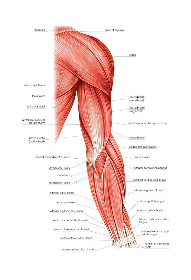
Muscles Of Right Upper Arm Photograph by Asklepios Medical ... from images.fineartamerica.com Attached to the bones of the skeletal system are about 700 named muscles that make up roughly half of a person's body weight. Muscular contraction is necessary for voluntary and involuntary movement of limbs, stabilization of joints, maintaining luminal diameter (in the case of arteries, bowel, etc), and to produce heat. • the muscular system develops from intra embryonic mesoderm. It allows the nervous system to trigger a specific movement of a muscle by. Each of these muscles is a discrete organ constructed of skeletal muscle tissue, blood vessels, tendons, and nerves. Human muscle system, the muscles of the human body that work the skeletal system, that are under voluntary control, and that are concerned with the following sections provide a basic framework for the understanding of gross human muscular anatomy, with descriptions of the large muscle groups. Find the best weight lifting exercises that target each muscle or groups of muscles. As the skeletal muscles pull on bones to cause movements, they also stabilize the joints of the skeleton;
Understanding the structure of a muscle fiber.
There's no strict demarcation or dividing line between the tendon and the covering around this muscle but that covering is called is called the epimysium fp my cm and it's really just connective tissue that covers the muscle kind of protects it reduces friction. This article will focus on tongue embryology, origin, insertion, and action of the extrinsic muscles, followed by innervation, blood supply and lymphatic drainage of the tongue. Skeletal muscles are attached to bones by tendons and can be as long as 30 cm, although they are usually 2 to 3 cm in length. Related online courses on physioplus. There are over two dozen gorgeous and painstakingly. See more ideas about muscle anatomy, anatomy, hip muscles anatomy. You can click the links in the image, or the links below the image to find out more information on any muscle group. Anatomy of the short head of the biceps brachii muscle. The primary function of the knee is to hinge at the lower extremity. Understanding the structure of a muscle fiber. There are two main muscle groups around the knee: Smooth muscles (involuntary muscles) are usually in sheets or layers, with one layer of muscle behind the other. Pulling the muscle refers to stretching the calf muscle beyond its limit.
More serious injuries may result in partial or complete tear of the calf. It allows the nervous system to trigger a specific movement of a muscle by. Skeletal muscles allow the body to move and maintain posture; The tendons of these muscles pass through a small corridor in the wrist known as the carpal tunnel. The muscular system consists of the skeletal muscles and their associated structures.
Muscles of the Anterior Leg - Attachments - Actions ... from teachmeanatomy.info This is a table of skeletal muscles of the human anatomy. Microscopic anatomy of skeletal muscle. Topographically, the muscles in this group are classed along with the lateral torso wall and upper extremity, which is due to their location as well as their genetic development based on their embryological origin. There are two main muscle groups around the knee: Inflammation of this region caused by repetitive stress or trauma may lead to pain and numbness known as carpal tunnel syndrome. Learning to draw muscles may conjure medical charts in daunting details, but such complexity is unnecessary. • definitions • introduction • development of muscles • classification • anatomy of skeletal muscle • muscle physiology • properties • muscles of development of muscles. You can click the links in the image, or the links below the image to find out more information on any muscle group.
• the muscular system develops from intra embryonic mesoderm.
See more ideas about muscle anatomy, anatomy, hip muscles anatomy. • definitions • introduction • development of muscles • classification • anatomy of skeletal muscle • muscle physiology • properties • muscles of development of muscles. Discover the muscle anatomy of every muscle group in the human body. As the skeletal muscles pull on bones to cause movements, they also stabilize the joints of the skeleton; Muscle mass accounts for a large majority of the carcass weight of domestic animals. As with muscles of other regions of the body, the various muscles of the upper and lower leg can be divided into groups. It allows the nervous system to trigger a specific movement of a muscle by. This article will focus on tongue embryology, origin, insertion, and action of the extrinsic muscles, followed by innervation, blood supply and lymphatic drainage of the tongue. There are over two dozen gorgeous and painstakingly. By contracting, they also aid the venous return of blood to the heart and with age, these components of the musculoskeletal system progressively degenerate, which contributes to frailty and increases the risk of falls and fractures. There are two main muscle groups around the knee: Attached to the bones of the skeletal system are about 700 named muscles that make up roughly half. Inflammation of this region caused by repetitive stress or trauma may lead to pain and numbness known as carpal tunnel syndrome.
Convergent muscles contain fibers that have a wide origin, but converge in order to attach to a narrow tendon. Find the best weight lifting exercises that target each muscle or groups of muscles. Learning to draw muscles may conjure medical charts in daunting details, but such complexity is unnecessary. It allows the nervous system to trigger a specific movement of a muscle by. The inguinal aponeurotic falx (falx aponeurotica inguinalis;
Anatomy Muscle Man Image - The Graphics Fairy from thegraphicsfairy.com Topographically, the muscles in this group are classed along with the lateral torso wall and upper extremity, which is due to their location as well as their genetic development based on their embryological origin. It allows the nervous system to trigger a specific movement of a muscle by. Muscle mass accounts for a large majority of the carcass weight of domestic animals. The muscle groups of the upper leg region are the gluteal group. Muscles of the thorax & abdomen | anatomy model. As the skeletal muscles pull on bones to cause movements, they also stabilize the joints of the skeleton; The anatomy of muscle cells differs from that of other body cells and biologists have applied specific terminology to different parts of these cells. The primary function of the knee is to hinge at the lower extremity.
Pulling the muscle refers to stretching the calf muscle beyond its limit.
The anatomy of muscle cells differs from that of other body cells and biologists have applied specific terminology to different parts of these cells. A collection of anatomy notes covering the key anatomy concepts that medical students need to learn. The muscles of the abdomen may be divided into two groups: Topographically, the muscles in this group are classed along with the lateral torso wall and upper extremity, which is due to their location as well as their genetic development based on their embryological origin. You can click the links in the image, or the links below the image to find out more information on any muscle group. Muscles of the upper and lower leg. The primary function of the knee is to hinge at the lower extremity. Learn about human anatomy muscles with free interactive flashcards. In this section, learn more about the anatomy of the muscles of the neck. Muscle movements, types, and names. Smooth muscles (involuntary muscles) are usually in sheets or layers, with one layer of muscle behind the other. As the skeletal muscles pull on bones to cause movements, they also stabilize the joints of the skeleton; Learn about the muscles, tendons, bones, and ligaments that comprise the knee joint anatomy.

